Valvular Heart Disease
• Left sided valve lesions:
– Aortic: stenosis / regurgitation
– Mitral: stenosis / regurgitation
• Right sided valve lesions:
– Tricuspid: stenosis / regurgitation
– Pulmonary: stenosis / regurgitation
• Prosthetic heart valves
Aortic stenosis
Aetiology
• Aortic stenosis may be congenital or acquired.
• Congenital malformations may be tricuspid, bicuspid or more rarely unicuspid / quadricuspid
• Acquired causes include the following:
- Degenerative disease
- Rheumatic disease
- Calcific e.g. end-stage renal failure, Paget’s disease
- Miscellaneous e.g. rheumatoid involvement
Pathophysiology

Symptoms
• Exertional dyspnoea or fatigue
• Angina
• Syncope
Physical findings
• Slow rising pulse
• Reduced systolic and pulse pressure
• Systolic thrill over the aortic area
• Ejection systolic, crescendo-decrescendo murmur
• Soft or inaudible second heart sound
• ECG: LVH, AV node conduction defects
Echocardiography
• Thickened valves with reduced motion, sometimes calcified
• Grading of stenosis severity is as follows:
- Normal valve area = 3-4cm2
- Mild stenosis = 1.5-3cm2
- Moderate stenosis = 1.0-1.5cm2
- Severe stenosis ≤ 1.0cm2
• When stenosis is severe, the peak gradient across the aortic valve is usually > 60mmHg.
Medical therapy
• Conservative treatment should be offered for mild to moderate aortic stenosis and to asymptomatic patients with severe aortic stenosis as follows:
- Advise to report symptoms
- Avoid vigorous exercise
- Antibiotic prophylaxis for endocarditis
- Regular follow-up ± echocardiography
Surgical/ Interventional therapy
• Aortic valve replacement should be offered to the following:
- Symptomatic pts with severe AS
- Pts with severe AS undergoing CABG surgery
- Pts with moderate AS undergoing CABG surgery
- Asymptomatic pts with severe AS and LV dysfunction
• Balloon valvuloplasty: bridge to surgery in haemodynamically unstable patients, or palliation for patients with serious comorbid conditions
• Transcatheter aortic valve replacement
Aortic valve replacement
• In the absence of LV dysfunction, operative risk is
2-5%.
• Indicators of higher mortality are NYHA class, LV dysfunction, age, concomitant coronary artery disease, and aortic regurgitation.
• Valve replacement usually results in reduced LV volumes, improved LV performance and regression of LV hypertrophy.
Transcatheter Aortic Valve (TAVI)
• Novel alternative therapy
• Performed via femoral, subclavian or transapical approaches
• Currently reserved for high risk, symptomatic severe degenerative aortic stenosis
Aortic regurgitation
Aetiology
• Either due to primary disease of the aortic valve or wall of the aortic root or both.
• Causes of primary aortic valve disease include:
- Congenital eg. bicuspid aortic valve
- Acquired: rheumatic valve disease, infective endocarditis, trauma, connective tissue disease.
• Causes of primary aortic root disease include:
- Degenerative, cystic medial necrosis (eg. Marfan’s), aortic dissection, syphilis, connective tissue disease, hypertension.
Clinical history
• Chronic severe AR
Dyspnoea is the principal symptom
Syncope is rare and angina is less frequent than in
aortic stenosis.
• Acute severe AR
LV decompensation occurs readily with fatigue,
severe dyspnoea and hypotension.
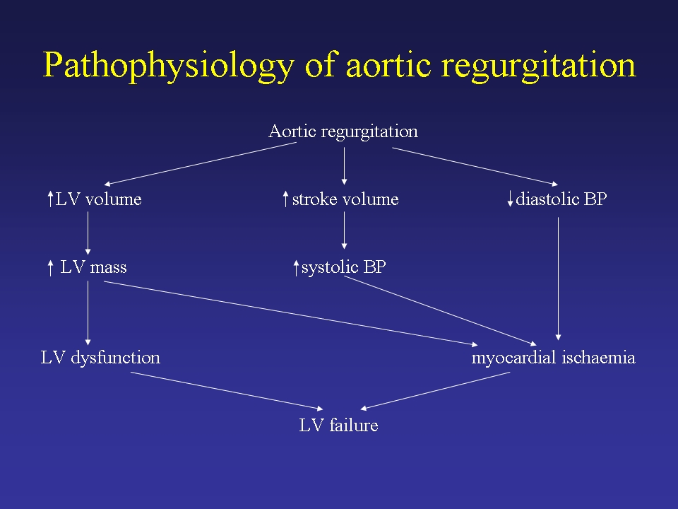
Pathophysiology of aortic regurgitation
Physical findings
• Collapsing pulse
• Wide pulse pressure
• Peripheral signs- De Musset’s, Corrigan’s, Quinke’s, Muller’s, Duroziez’s
• Hyperdynamic apex beat
• Early blowing diastolic murmur
• ECG: Left axis deviation, LV hypertrophy.
• CXR: Cardiomegaly, aortic calcification, aortic root dilatation
Echocardiography
• Colour flow
- width of the jet at its origin
- extent into the LV
• Doppler
- Rate of decline of aortic reguritant flow
- Diastolic flow reversal into the descending aorta
Management
• Medical treatment
- Diuretics, digoxin, salt restriction
- Vasodilators
- Endocarditis prophylaxis
• Without surgery, death usually occurs within 4 years of developing angina and within 2 years after onset of heart failure.
Surgical therapy
• Severe acute AR requires prompt surgical intervention.
• Chronic severe AR
- Symptomatic patients with normal LV function
- Symptomatic patients with LV dysfunction or dilatation
- Asymptomatic patients with LV dysfunction or dilatation (EF<50% or end-systolic diameter > 55mm)
• Aortic valve and root replacement- if aortic root diameter is ≥ 50mm.
Mitral stenosis
Aetiology
• Rheumatic
• Congenital
• Carcinoid, SLE, rheumatoid arthritis, mucopolysaccharidoses.
• Left atrial myxoma, ball-valve thrombus, infective endocarditis with large vegetation and cor triatriatum.
Rheumatic mitral stenosis
• Fusion of the valves, commisures and chordae
• Symptoms usuually occur in the 3rd or 4th decade, but mild MS in the aged is becoming more common.
• 25% of patients have pure mitral stenosis and two-thirds are female.
• Lutembacher’s syndrome- associated with an atrial septal defect
Pathophysiology
• Normal mitral valve area = 4-6cm2.
• A mitral valve area ≤ 1cm2 equates to severe mitral stenosis.
• Symptoms usually develop when mitral valve area ≤ 2.5cm2
• Symptoms in mild mitral stenosis usually precipitated by exercise, emotional stress, infection, pregnancy or fast atrial fibrillation.
Natural history
• Long latent period of 20 to 40 years
• Once significant limiting symptoms occur, 10-year survival rate is 5-15%.
• With severe pulmonary hypertension, mean survival falls to
< 3 years.
• Mortality from untreated mitral stenosis is due to progressive heart failure (60-70%), systemic embolism (20-30%) and pulmonary embolism (10%).
Clinical features
• Dyspnoea
• Haemoptysis may also occur
• Angina
• Embolic events
Physical findings
• Mitral facies
• Tapping apex beat
• Right ventricular heave, loud P2
• Loud first heart sound.
• Opening snap.
• Rumbling, mid-diastolic murmur with presystolic accentuation in sinus rhythm.
Echo evaluation
• Assessment of valve morphology: degree of leaflet thickness, mobility and calcification and extent of subvalvular fusion.
• Estimation of left atrial size.
• Doppler echo: estimation of mitral valve area, transvalvular gradient and PA pressure.
Medical treatment
• The asymptomatic patient with mild mitral stenosis should be managed medically. Medical therapy includes:
- Avoidance of unusual physical stress.
- Salt restriction.
- Diuretics if needed.
- Control of heart rate – β-blocker or digoxin.
- Anticoagulation for AF or prior embolic event.
- Annual follow-up.
- Echocardiography if deterioration in clinical condition.
Management of symptomatic mitral stenosis
• Patients with symptoms should undergo clinical re-evaluation with echocardiography.
• NYHA class II symptoms and mild mitral stenosis may be managed medically.
• NYHA class II symptoms and at least moderate stenosis (MVA≤1.5cm2 or mean gradient ≥5mmHg) may be considered for balloon valvuloplasty.
• NYHA class III or IV symptoms and severe mitral stenosis should be considered for balloon valvuloplasty or surgery.
Mitral valve replacement
• Severe mitral stenosis and contraindications to surgical commisurotomy or balloon valvuloplasty:
- Restenosis following surgical commisurotomy or balloon valvuloplasty
- Significant mitral regurgitation
- Extensive calcification of the subvalvular apparatus.
• Operative mortality ranges from 3-8% in most centres.
Mitral Regurgitation
Chronic MR
Acute MR
Mitral valve prolapse
Aetiology
Mitral regurgitation may be caused by abnormalities of the valve leaflets, chordae tendinae, papillary muscles or mitral annulus:
• Valve leaflets
- myxomatous degeneration
- rheumatic heart disease
- infective endocarditis
• Chordae tendinae
- congenital, infective endocarditis, trauma, rheumatic fever, myxomatous
• Papillary muscles
- myocardial ischaemia, congenital abnormalities, infiltrative disease
• Mitral annulus
- dilatation eg. ischaemic or dilated cardiomyopathy
- calcification due to degeneration, hypertension, diabetes,
chronic renal failure
Clinical features
• Symptoms usually occur with LV decompensation: dyspnoea and fatigue.
• Physical findings include:
- Pulse: sharp upstroke
- Apex: displaced, hyperdynamic
- Pansystolic murmur
Natural history
• The natural history of chronic MR depends on the volume of regurgitation, the state of the myocardium and the underlying cause.
• Preoperative LV end-systolic diameter is a useful predictor of postoperative survival in chronic MR.
• The preoperative LV end-systolic diameter should be < 45mm to ensure normal postoperative LV function.
Medical treatment
• Symptomatic patients may benefit from the following drug therapy whilst awaiting surgery:
• Vasodilator therapy
• Diuretics
• Digoxin / Beta-blockers in presence of atrial fibrillation.
• Endocarditis prophylaxis
Surgical treatment
• Symptoms or left ventricular end systolic diameter ≥45mm.
• Mitral valve repair or replacement.
• Mitral valve repair better preserves LV function and avoids the need for chronic anticoagulation.
Acute mitral regurgitation
Aetiology
Important causes of acute mitral regurgitation include:
• Infective endocarditis
• Ischaemic dysfunction or rupture of papillary muscle.
• Malfunction of prosthetic valve.
Chronic versus Acute MR
Finding Chronic MR Acute MR
Symptoms subtle onset obvious
Appearance normal/mildly severely ill
dyspnoeic
Tachycardia not striking always present
Apex beat displaced not displaced
Systolic thrill common a bsent
Murmur harsh pansystolic soft or absent early systolic component
ECG-LVH usually present absent
CXR severe cardiomegaly normal heart size
Acute mitral regurgitation
Medical therapy
The following therapies may be beneficial in reducing the severity of MR
- Vasodilator therapy
- Inotropic therapy
- Intra-aortic balloon counterpulsation
Surgical therapy
• Indicated in patients with acute severe MR and heart
failure.
• Higher mortality rates than for elective chronic MR
Mitral valve prolapse
General features
• 2-6% of the general population
• Twice as common in women.
• Due to myxomatous proliferation of the mitral valve.
• Primary condition, or secondary finding in connective tissue
diseases e.g. Marfan’s syndrome.
• Vast majority asymptomatic.
• Palpitations, dizziness, syncope, or chest discomfort.
• Mid-systolic click, late systolic murmur
Echocardiographic criteria
• M-mode criterion: ³ 2mm posterior displacement of one or both
leaflets.
• 2-D echo findings: Systolic displacement of one or both leaflets
within the left atrium in the parasternal long-axis view; leaflet
thickening, redundancy, chordal elongation and annular dilatation.
Natural history
• Benign prognosis in most patients
• Complications may occur in patients with a systolic murmur,
thickened leaflets, an increased LV or LA size, especially in men
> 45 years old
• Complications include progressive mitral regurgitation, infective
endocarditis, cerebral emboli, arrhythmias and rarely sudden death.
Management
• Asymptomatic patients without MR or arrhythmias have an excellent prognosis – follow-up every 3-5 years.
• Patients with a long systolic murmur may show progression of MR and should be reviewed annually.
• Severe MR requires surgery, often mitral valve repair.
Tricuspid stenosis
• Almost always rheumatic
• The low cardiac output state causes fatigue; abdominal discomfort may occur due to hepatomegaly and ascites.
• The diastolic murmur of tricuspid stenosis is augmented by inspiration.
• Medical management includes salt restriction and diuretics.
• Surgical treatment in patients with a valve area <2.0cm2 and a mean pressure gradient >5mmHg.
Tricuspid regurgitation
• Most common cause is annular dilatation due to RV failure of any cause
• Symptoms and signs result from a reduced cardiac output, ascites, painful congestive hepatomegaly and oedema.
• The pansystolic murmur of TR is usually loudest at the left sternal edge and augmented by deep inspiration.
• Severe functional TR may be treated by annuloplasty or valve replacement. Severe TR due to intrinsic tricuspid valve disease requires valve replacement.
Pulmonary stenosis
• Most commonly due to congenital malformation
• Survival into adulthood is the rule, infective endocarditis is a risk and right ventricular failure is the most common cause of death.
• Carcinoid plaques may lead to constriction of the pulmonary valve ring.
Pulmonary regurgitation
• Most common cause is ring dilatation due to pulmonary hypertension, or dilatation of the pulmonary artery secondary to a connective tissue disorder.
• May be present and well-tolerated for many years
• The clinical manifestations of the primary disease tend to overshadow the pulmonary regurgitation.
• Physical examination
– right ventricular heave
– high-pitched, blowing, early diastolic decrescendo murmur
• left sternal edge
• augmented by deep inspiration.
• Pulmonary regurgitation is seldom severe enough to require specific treatment. Surgery may be required because of intractable RV failure.
Prosthetic valves
Prosthetic valves may be divided into 2 broad categories:
Mechanical valves
• Very good durability.
• Require long-term anticoagulation.
• May cause mild haemolysis.
Bioprosthetic (tissue) valves
• Porcine variety most commonly used.
• Limited durability.
• Anticoagulation for first 3 months only.
Mechanical versus tissue valves
• No difference in survival, haemodynamics or in probability of
developing endocarditis, valve thrombosis or systemic embolism.
• Valve-related failure is much more common with tissue valves.
• Anticoagulant-related bleeding occurs with mechanical valves.
• Elderly patients tend to receive tissue valves.
Satheesh Nair
Manchester Heart Centre


Good post.Valuable electure notes..for simpson's Forensic medicine book free download visit : http://dentistryandmedicine.blogspot.com/2011/06/free-download-free-simpsons-forensic.html
ReplyDeleteDIS IS AWESME!!!VERY USEFUL FOR QUICK REFERRAL WEN NOT HAVIN THE TEXTBUK
ReplyDelete